Outpatient acute seizure management at the level of the general practitioner clinic: a proposed treatment algorithm

Outpatient acute seizure management at the level of the general practitioner clinic: a proposed treatment algorithm


Prahlad K Sethi, MD1 Dhrumil Shah, MD1 Anuradha Batra, MD 1 Nitin K Sethi, MD2


1 Department Neurology, Sir Ganga Ram Hospital, New Delhi, India

2 Department of Neurology, New York-Presbyterian Hospital, Weill Cornell Medical Center, New York, NY (U.S.A.)


Seizures beget seizures has been a point of contention over the years. There is some scientific evidence to suggest that each seizure increases the risk for future seizures and that failure to control seizures in a timely fashion can lead to status epilepticus (SE). Status epilepticus is a life threatening neurological emergency which can present as an exacerbation of a pre-existing seizure disorder such as in an epilepsy patient who is non-compliant with his anti-epileptic drug (AED) regimen or as the initial manifestation of a seizure disorder (epilepsy) or as the manifestation of other systemic and cerebral insults. Prolonged seizures are also associated with worse neurological outcomes. With the aim to reduce the time to treatment gap, outpatient treatment of seizures is now been explored. We discuss this approach in relation to the health care system of India.


General practitioners (GPs) also referred to as primary care physicians (PCPs) form the backbone of the Indian health care delivery system. Usually, they are in solo practice working in small clinics (offices) which are ill-equipped to handle medical and surgical emergencies such as seizures and SE.

As awareness about coronary artery disease has increased, GPs now feel comfortable administering aspirin and nitrates before transferring the patient to the hospital. With respect to emergency management of seizures, their knowledge and experience is more limited. If the seizure has stopped, the patient is usually referred to a neurologist. If the patient is actively seizing, the patient is referred to the nearest hospital. The time to treatment gap results in increased morbidity and mortality especially for patients presenting with SE. Unlike developed countries, India lacks a well-organized and responsive 911 type medical emergency system manned by well trained and certified emergency medical technicians (EMTs) and paramedics who can administer life-saving 1st line and 2nd line antiseizure medications such as benzodiazepines (lorazepam, diazepam) and phenytoin or fosphenytoin parenterally en-route to the hospital.


Status epilepticus is a neurological emergency. Early effective treatment of SE results in termination of seizure activity and thereby reduction in cerebral hypoxia and damage. Hence every effort should be made to treat SE at the earliest. In India this goal would be best achieved by initiation of treatment of SE at the GP level. Till recently only intravenous benzodiazepine (diazepam or lorazepam) or rectal diazepam was available for the emergent management of seizures and SE. Now drugs which can be rapidly administered via the intranasal or intramuscular routes are also available such as intranasal midazolam and intramuscular fosphenytoin. Neurocritical Care Society guidelines recommend the administration of benzodiazepines via rectal, intramuscular, intranasal or buccal routes if intravenous or oral administration is not feasible. 1Administration via the above routes has been determined to be quick, easy, safe and to achieve high and consistent blood levels of the active compound. 2


We feel that antiseizure drugs in these formulations can be easily administered by a GP at the clinic without any special expertise or formal training.


Intranasal midazolam: Currently in Indian market 2 midazolam formulations are available, one is MIDACIP (Cipla Pharmaceuticals) and other is MIDASPRAY (Intas Pharmaceuticals). They are both metered spray preparations. Two strengths of MIDACIP are commercially available: (1.25mg and 0.5mg)


Method of administration of MIDACIP nasal spray (Figure 1):


  1. Shake the bottle gently.
  2. Remove the dust cap.
  3. Hold the bottle with your forefinger and middle finger on either side of the nozzle and your thumb underneath the bottle
  4. If using first time, spray it six times in the air with the nozzle pointing away from the patient until the consistent mist of the drug is delivered, this is called priming, which ensures that correct dose is delivered.
  5. If the patient is in supine position, head is slightly lifted upwards and the device should be placed near the patient’s nose.
  6. Insert the nozzle into patient’s nostril, depress the pump with a firm even stroke. (Patient need not inhale)
  7. Tilt the patient head backward while spraying, this will avoid swallowing of the solution.
  8. Administer one spray at a time in each nostril to continue prescribed dose
  9. Reprime the device for subsequent use if the bottle is not used for more than a day. To reprime spray it two to three times in the air until a fine mist appears. For reusing the device nozzle and dust cap must be washed before storage.


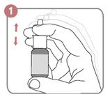
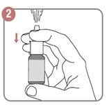
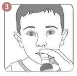
Figure 1. Method of administration of intranasal midazolam.


Each nasal spray delivers either 1.25mg or 0.5mg of midazolam. Dose is titrated according to individual patient weight and full effective dose should be administered. For adults, dose is 5 mg if weight <50 kg and 10 mg if weight>50 kg. The dose should be equally divided and administered into each nostril. For children the recommended dose of MIDACIP nasal spray is 0.2 mg/kg body weight. The dose should be equally divided and administered into each nostril. Placing half the medication in each nostril reduces the volume while doubling the available surface area for absorption.


Table 1. Dosing Guidelines of MIDACIP Nasal Spray in children


Age (years) Weight (kg) Dose (mg) Metered Doses in Each Nostril
½ – 1 <10 1.25 – 2 1 – 2
1 – 4 10 – 16 2.5 2 – 3
4 – 10 16 – 32 5 4 – 6
>10 > 32 10 10


Intramuscular midazolam and fosphenytoin: GPs by virtue of their training can administer intramuscular injections with ease. In India intramuscular midazolam (MIDAZ, Abbott Healthcare or FULSED, Ranbaxy Laboratories) injections are widely available and cost effective. Midazolam is given intramuscularly at the dose of 10mg once or 0.2mg/kg once but not exceeding 10mg. Intramuscular fosphenytoin formulations are also available (Fosolin, Zydus Cadila Healthcare or Fosphen, Intas Pharmaceuticals) but more expensive. These preparations are available as ampoules of 150mg (75mg/ml, 2ml). It is our recommendation that the GP administer 2 ampoules of fosphenytoin stat in the clinic prior to transporting the patient to the hospital. Doing so may abort the seizure and possibly terminate the SE.


Use of the above formulations in the clinic setting by the GP along with established seizure first aid guidelines (Figure 2) has the potential to save many lives and reduce the morbidity from seizures and SE in our country.


Figure 2. Seizure first-aid guidelines (Source: Epilepsy Foundation Eastern Pennsylvania, www.epepa.org)


References


  1. Brophy GM, Bell R, Claassen J, et al. Guidelines for the evaluation and management of status epilepticus. Neurocrit Care 2012; 17:3–23.


  1. Agarwal SK, Cloyd JC. Development of benzodiazepines for out-of-hospital management of seizure emergencies. Neurol Clin Pract. 2015; 5:80-85.


Side-effects associated with anticonvulsant use

Anticonvulsants (anti seizure medications) use is associated with various side-effects. Some of these can be troublesome. In this post I shall briefly list a few of the side-effects common to a number of anticonvulsants. As always my advise remains unchanged. The information provided here is no substitute to an actual visit to your physician. But I hope this post shall make you better informed.

Rash: can be associated with the use of a number of anticonvulsants. Phenytoin, lamotrigine, carabamazepine are the anticonvulsants commonly associated with rash. The rash may be mild or it may become quite fulminant leading to involvement of the mucous membranes (Steven Johnson Syndrome). The rash usually appears right at the onset (meaning a few days after the medication is started) but it may also appear at any point of time during the course of therapy. If rash is documented the usual advise is to stop the medication and consult your physician as soon as possible. Please remember though that sudden stoppage of anticonvulsant is not advisable since it may lead to a flurry of seizures. So it is your doctor who shall be the best person to make the decision: either stopping the medication cold turkey and substituting another anticonvulsant in its place  or slowing down the upward taper of the anticonvulsant and allowing the rash to subside.

Cognitive side-effects: a number of anticonvulsants can cause cognitive side-effects. Patients may complain of feeling dull (“I do not feel as sharp as usual” or “my mind is in a fog”). Some complain of difficulty concentrating and focusing while others have word finding difficulty (has been reported with the use of topiramate). Again if you experience any of these side-effects bring them to the attention of your doctor. At times lowering the dose of the anticonvulsant leads to resolution of these side-effects. At times taking the bulk of the anticonvulsant at night (larger dose at night and smaller dose in the morning) may be helpful since you can sleep off most of the side-effects.

Bone loss: many anticonvulsants lead to bone loss. The most commonly cited culprit drugs are phenytoin, carbamazepine and phenobarbital. Prolonged use of these anticonvulsants leads to bone loss and osteoporosis. That is the reason why your physician may advise you to supplement calcium and vitamin D. How much calcium and vitamin D to take on a daily basis though? The National Osteoporosis Foundation and National Institute of Health has given recommendations for daily calcium and vitamin D intake and most of the physicians refer to these.  Again your physician shall be the best person to determine how much calcium and vitamin D supplementation is required based on your age, the various medications you are taking and your risk of osteoporosis in the future. He may refer you for a bone densitometry test (commonly referred to as a DEXA scan).

Congenital malformations (major and minor): this applies to women of child bearing age who are exposed to/ taking anticonvulsants. Data from various pregnancy registries collected over many years has now informed us that anticonvulsant use by a woman during pregnancy may at times lead to major or minor malformations (cleft lip, cleft palate, congenital heart disease, spina bifida and so forth) in the baby. The risk though varies with some anticonvulsants “safer” than others.  No anticonvulsant though is completely free of this risk and again it is your doctor who shall be the best person to advise you about this.  The choice of an anticonvulsant in a woman of child bearing age is made after due consideration of the above risk. Folic acid supplementation before and during pregnancy may help to mitigate some of this risk to some extent.  So it is imperative that all women of child bearing age who are on anticonvulsants (and are contemplating pregnancy) should have a discussion about the risk of congenital malformations with their doctors.

Mood changes: anticonvulsants can lead to mood changes. Studies have shown that some are more likely to do so than others. Patients may complain of low mood, caregivers may notice that the patient is more agitated, snappy or restless. Some patients may become depressed. Hence the FDA has issued a warning on anticonvulsants and the risk of suicide and suicidal thoughts. Again your physician shall be the best person to decide which anticonvulsant is appropriate for you.

Apart from the above mentioned side-effects, each anticonvulsant has side-effects which are unique to it.  So it is important that you read the package insert and tell your doctor about any other medical conditions that you may have. Also mention other medications that you taking so that your doctor can determine and tell you about important drug-drug interactions.

I hope this information helps some of the readers of my blog.


Nitin Sethi, MD

A question from a father and some answers for seizures due to hypoglycemia

A father from the Netherlands recently wrote to me about his son. I thank him for his question.  He asked me a few very specific questions. I am reproducing them here as I feel it shall aid other people. My response to his questions follows. Names have been removed to maintain privacy.

QUESTION:

My son is diabetic type 1, since his 5th year. And he had several seizures in the last 5 years. Mostly once a year, every time he had a hypoglycaemia.
The last time he had a epileptic seizure, with a fracture of thoracic vertebra 2. I have made a MRI scan of the brains, but everything seems normal.
Are there other investigations necessary to be sure it is an epileptic insult due to hypoglycaemia, instead of real epilepsy

HK

ANSWER:

Dear HK,

                                  thank you for writing in to me. If I get you right you are saying that all of your son’s seizures have occured in the setting of hypoglycemia. Do you by any change recall what was his blood sugar during these ictal episodes. For a seizure to occur due to hypoglycemia, the blood sugar has to usually fall down to an extremely low level (we call this neurohypoglycemia). As you are aware the brain needs sugar for energy and its metabolism so when blood sugar falls down to the range of 60 mg/dl or less, that is when a seizure occurs. The seizure in the setting of hypoglycemia is usually a generalized convulsion. That said I and many other physicians have seen patients presenting to the ER with focal findings suggestive of a stroke only to find them completely reverse once the blood sugar was corrected.

I do have a few practical suggestions for you:

–have a home blood glucose meter and check your son’s blood sugar at various times during the day such as in the early morning when he wakes up (fasting blood sugar), prior and after lunch (post prandial blood sugar) and finally before he heads to bed during the night.  Do this for about 2 weeks and keep a record of the values in a notebook. This shall prove to be tremendously helpful to you as a parent and his physician to see how his blood sugar fluctuates during the day. Are there times when his blood sugar falls down/ bottoms out unexpectedly.

–a seizure due to hypoglycemia is rapidly reversible and in fact can be aborted with intravenous dextrose solution. Now it may be hard to administer intravenous dextrose at home.

–another very important point which comes to mind is why is he having so frequent hypoglycemic episodes. This shall require a thorough review by his endocrinologist. His insulin dose may need to be adjusted and/or he may warrant frequent small meals during the day to prevent his sugar from suddenly dropping.

–now to your final question: does your son actually have underlying epilepsy per-se. The answer to that depends upon this:

                -has he ever had a seizure in the setting of normal glucose level?

                -an EEG study shall be extremely useful. If the electroencephalogram shows inter-ictal epileptiform activity then likely your son does suffer from epilepsy. Remember in patients with seizures due to hypoglycemia per-se, the EEG between the seizures should be normal.

                -a routine EEG (30 minutes study) can fail to reveal an answer. In that case a long term EEG monitoring study (24-72 hours either in the hospital or in an outpatient setting) may aid in characterization of his typical events.

-as you may have guessed right, patients with seizures due to hypoglycemia per se do not need to be on anti-convulsant therapy. In these patients what we need to ensure that they do not become hypoglycemic. No hypoglycemia means no seizures.  On the other hand if your son does have underlying epilepsy, he shall warrant anti-convulsant therapy.

                -also a neuroimaging study (ideally MRI of the brain) may he helpful to rule out any intracerebral structural cause of seizure.

I hope you find this information helpful. Please feel free to contact me again.

Personal Regards,

Nitin Sethi, MD

Syncope Vs Seizure: the quest for an answer

One of the readers of my blog sent me an interesting query. Here is her history. My response to it follows. I have deleted her name and hidden her email address to maintain her confidentiality.


HISTORY

Hi! I was grocery shopping in Wal-mart when I had my episode. I was walking to the front of the store after shopping for about an hour. My vision kept blacking out. It was like someone just turned out the lights. I would be able to see again and I would be standing there staring at something. This happened about 4 or 5 times over a ten minute span. The next thing I knew I was waking up in the hospital. Apparently I hit the ground started convulsing, my eyes rolled back and I was foaming at the mouth. I bit the side of my tongue, my body was horribly sore the next couple of days, but I didn’t have any bladder issues. I had no memory of the seizure or what had happened. I kept going in and hour for the next couple of hours. I busted my head on the floor which required staples. I had memory problems and balance issues for the next week. I then made an appointment with a neurologist. He thinks I have syncope. He sent me for my EEG to rule out epilepsy, but we haven’t got the results back yet. While I was having my EEG I started having facial spasms during the flashing light portion of the test. I’m reading about syncope and some of it just doesn’t fit. I am a HUGE salt eater. I crave salty foods all day! So if that was it, wouldn’t my diet be treating the syncope? Also, my sister has epilepsy. Her seizures are triggered by flashing lights. I have never had a seizure before. I have passed out in the past, but it was because of hypoglycemia. I don’t have any issues with heat or pain or seeing blood. It just doesn’t affect me. I just don’t know if he’s got the correct diagnosis and would like someone else’s opinion. Thanks!

Dear A,
                 thank you for writing in to me.  Your history has intrigued me and hence I shall dwell over it a little before offering my humble opinion.  It goes without saying, this opinion is offered without taking your history in person and examining you.  You should follow what your doctor tells you.  He/ she shall be the best person to guide further diagnostic and management issues.  Your recent event had features of both syncope as well as a seizure/ convulsion. So what in your history points towards syncope?
PRODROMAL FEATURES: meaning things which you felt prior to passing out. You felt light headed, your vision was blurred/ tunneled/ kept going in and out (it always fascinates me how patients use different words to describe the same symptom). Prior to a syncopal event patients may feel as if they are about to pass out/ faint. They may look pale (all the blood was drained out from the face), they may feel/ complain that their legs feel weak/ woobly.
I am uncertain what to make of the staring episodes which followed soon after. By staring do you mean you were unable to concentrate (may occur with syncope) or do you mean you had impairment in your level of consciousness and awareness ( goes more towards a seizure).
Anyways let us move forward. The next thing you remember is waking up in the hospital. From the bystander history, you were noticed to have convulsive movements. During the seizure, you lost body tone leading to a hard fall which cracked open your skull.  During the convulsion itself your eyes rolled back into your head, you foamed from the side of the mouth and bit the side of your tongue. Yes biting the side of the tongue goes more towards a seizure than biting the tip of your tongue. I am not sure who figured that one out though. You did not have loss of bladder control. Post the seizure, you were not yourself  for the next few days with a slow return to the baseline.
There I think I have summarized your history well. Well what happens next? Your history has features of both syncope as well a seizure.  I would have asked a few more questions:
1) did you feel anything prior to the event. We call these auras. Any strange smell, any strange taste and so forth.
2) have you ever had a seizure before.
3) any history of febrile convulsions?
4) are you prone to syncope: were you dehydrated, sick with the flu and so forth.
Well let us move forward. I am taking you step by step as I work through this history. So the doctor ordered an EEG (test to look at the brain waves). Why the EEG? Well simple if the EEG shows abnormal brain waves (I use the word misfiring of the brain), it points towards a seizure. If the EEG is normal, it may point towards syncope. That said and done, patients with seizures may have a normal EEG.
We do not have the EEG results. But during the test you mention something happened to you. Your face started twitching while photic stimulation was been carried out (flashing lights). Your sister has epilepsy and you say her seizures are triggered by flashing lights (usually patients who have primary generalized epilepsy have these kind of seizures).
So where does all this lead to?  My opinion: it is possible you have an underlying seizure disorder.
My recommendations: I would try my level best to rule out or rule in seizures. This may need a longer duration EEG study, if the first one is unrevealing. The decision to start anti-convulsant therapy shall be guided by all the above : history, examination findings, EEG findings and neuroimaging findings (CT scan or MRI brain).
THE BEST PERSON TO MAKE THAT DECISION–YOUR CURRENT DOCTOR AND NOT ME OVER THE INTERNET.  

Post traumatic epilepsy: a question and an answer

One of the readers of my blog asked me a question regarding her son. I am reproducing her question here. My answer to it follows.

V

my son was involved in a car crash 3 years ago aged 17 years and required brain surgery to remove a bloodclot. He recovered well with no ill effects. He had his first fit in November 2009. He had another 2 that same month. CT and MRI scans clear but eeg showed slight abnomal waves over area of surgery. Neuroligist said our choice if wanted to be on medication. Last fit was 27th November 2009 but he has just had another on 8th February whilst flying to holiday Do you thnk meds should be started to prevent further fits.

Dear V,

                    thank you for writing in to me. Your son’s condition is consistent with what is called post traumatic epilepsy. Let me attempt to explain this a little further. Your son obviously was not born with a seizure disorder (epilepsy). He was apparently well till he was involved in a motor vehicle accident (MVA). As a result of the MVA, he suffered head injury and from what you decribe an intracranial hematoma (blood clot) which required evacuation (removal of the blood clot surgically). He had an uneventful recovery but soon there-after had his first convulsion (you do not mention the time interval between the head trauma and the first convulsion).

As the name suggests post traumatic epilepsy refers to epilepsy/ seizure disorder which occurs after head trauma. Usually for post traumatic epilepsy to occur, the head trauma has to be significant such as a motor vehicle accident with significant intracranial hemorrhage or head injuries sustained in the battle field. Many of our soldiers returning from the battlefields of Afghanistan and Iraq suffer bullet shot injuries to the head (these as you can imagine are penetrating head injuries and cause significant brain damage as the high velocity bullet traverses through the skull). IED (improvised explosive devices) related blast injuries cause closed but still significant head trauma and are the signature injury of these two wars. Many of these brave men and women later develop post traumatic seizure disorder/ epilepsy. In other words minor bumps to the head (example you walk into a door) do not cause post-traumatic epilepsy.

There are three types of post traumatic epilepsy. Immediate, early and delayed. Let me explain this at length. Let us assume you are involved in an accident. Your head strikes the ground or steering wheel hard. You have a seizure soon after the impact. This is called immediate or impact seizure. This type of seizure does not lead to seizures later in life and hence such a patient does not warrant to be on long term anti-convulsant therapy.

Early post traumatic seizures are those which occur within 6 months of injury while late post traumatic seizures are those which occur after 6 months. Remember you can have your first post traumatic seizure as long as 5 years after the head injury. In other words if 18 months go by and the person has not had a seizure then likely he shall not have seizures as a result of head trauma. Patients who have early and late post traumatic epilepsy may warrant treatment with anti-convulsants. This is because the brain has suffered a scar (as a result of the head injury) and it is this scar tissue (consisting of damaged brain tissue) which then misfires and acts as a seizure focus (point in the brain where the seizure originates from).

In the case of your son, since he has suffered multiple convulsions since his head injury, he likely needs to be on an anti-convulsant. This decision though shall be made by his neurologist after consideration of factors which I mentioned earlier in my post. EEG may or may not be helpful in this regard (a normal EEG does not rule out seizure disorder and vice versa not every patient with a seizure disorder has an abnormal EEG).

I hope this is helpful to you. I wish him my very best.

Personal Regards,

Nitin Sethi, MD

Post traumatic epilepsy or seizures after head trauma

Post Traumatic Epilepsy

Nitin K Sethi, MD

Assistant Professor of Neurology

New York-Presbyterian Hospital

Weill Cornell Medical Center

New York, NY 10065


In this post I shall discuss the entity called post traumatic epilepsy/ post traumatic seizure disorder.  Epilepsy is a condition characterized by two or more seizures in a person’s lifetime. Broadly speaking epilepsy can be of two kinds:

1. Primary Epilepsy

2. Secondary Epilepsy

Patients who have primary epilepsy have seizures usually due to an underlying genetic predisposition. They do not have a secondary cause for their seizures and neuroimaging is usually normal.  On the other hands patients who have secondary epilepsy usually have seizures secondary to something (example secondary to brain tumor, secondary to an abscess in the brain, after a stroke and so forth). Under this category of secondary epilepsies is included post traumatic epilepsy (as the name suggests patients have seizures secondary to brain trauma).

Let me explain with the aid of an example.  Let us assume our patient (we shall call him Philip) is a 27-year-old healthy male with no significant medical or surgical history. Bikes are his passion especially Harleys.  Have you seen the ones they show on American Chopper. But we are digressing from our story line. Philip loves to ride them fast. A bright sunny Sunday morning finds him zipping down FDR drive at 80 mph.  With a bike under me, I felt like a real man. And then disaster strikes. Philip’s bike gets clipped by a speeding SUV. Philip is flung from the bike and hits the ground hard. Did I mention he was not wearing a helmet at this time. He is rushed to the nearest hospital. A lacerated spleen, couple of broken ribs and a fractured collar bone. Not too bad you might say. He shall live to ride another day. But all is not so rosy. Philip does not regain consciousness and does not respond to verbal commands.  A quick CT scan yields the answer. Philip has suffered extensive bleeding in the brain (neuro trauma). He is admitted to the neurological ICU. Recovery is painfully slow and after a months stay in the hospital, Philip is discharged to a sub-acute rehab facility. Alls well that ends well? Not quite done yet, I am afraid. Six months after his motorbike accident, Philip is again rushed back to the hospital after a witnessed tonic clonic convulsion. He is evaluated by a neurologist (like me) and a diagnosis of post traumatic epilepsy is made.

So what exactly is post traumatic epilepsy? As the name suggests epilepsy develops after head injury. Seizures can occur anytime after head injury. If they occur immediately after head injury it is referred to as immediate post traumatic epilepsy (also called impact seizures, as seizures occur at the time of impact to head).  If seizures occur within the first month after head injury it is referred to as early post traumatic epilepsy. Patients may have their first seizure as long as 18 months after head trauma. This is referred to as late post traumatic epilepsy.

Patients develop post traumatic epilepsy as a result of scarring of brain tissue.  They usually have convulsions. The treatment of post traumatic epilepsy is essentially the same as that of any other type of epilepsy. Once the seizure type is characterized, the right anti-epileptic drug is usually effective in controlling the seizures.

Is it a seizure or is it syncope? the story continues….

                      Is it a seizure or is it syncope? the story continues….

So our story ended with John in the ER. As many of you rightly guessed the first case scenario represents a typical syncopal episode while in the second case John had a generalized convulsion (seizure).

So what are the points in the history which favor syncope and which favor a seizure?

When a patient presents to a neurologist with an episode of loss of consciousness, it is imperative that we try to elucidate the underlying cause. As you can imagine the treatment of both these conditions is very different.

Syncope (fainting) can come either from the heart (we call this cardiogenic syncope) or from the brain (we call this neurogenic syncope or vasodepressor syncope or more commonly as vasovagal syncope). So for example you can faint (have a syncopal episode) if you have a sudden massive heart attack, or a transient arrhythmia of the heart (the heart beat fluctuates). As you can imagine these are potential lethal causes and hence patient’s who present with syncope are frequently evaluated for these cardiac conditions. Tests like ECG, prolonged 24 ECG (electrocardiogram) and sometimes an echocardiogram are ordered. Vasovagal syncope on the other hand is more benign and our patient John likely had a vasovagal syncopal episode in case scenario No 1. Another classical example of vasovagal syncope is when someone faints when he or she sees blood for the first time (frequently reported in medical students when they go into the OR for the first time).

 So what are the points which favor syncope?

1. Feeling light-headed prior to the episode

2. Feeling dizzy as if you are about to faint.

3. Blurring of vision at the onset of the episode ( Doctor I felt light headed, a little woosy, my vision started to go black and then I passed out)

4. Syncope usually occurs in an upright position (patient is usually standing when it occurs). Syncopal patients usually do not shake (that is they do not have convulsive movements. There is an entity called syncopal convulsion where in the episode starts with a syncope but then goes on to become a seizure. I shall not go into the details here as then it shall become confusing).

5. Usually the loss of consciousness is of very short duration. Once they fall to the ground and the blood rushes to their brain (as gravity has been eliminated), they rapidly regain consciousness.

6. They are not confused after the episode. They come around rapidly and know where they are (they are not confused and disoriented after the episode).

7. Syncopal patients usually do not bite their tongue or have loss of bladder control (wet their patients) during an episode.

What are the points which favor a seizure?

1. Patients who have a seizure do not get the type of prodomal symptoms which patients with syncope do. Meaning they do not feel light-headed, dizzy as if they are about to pass out. Seizures frequently occur out of the blue with no warning whatsoever. That said and done, some patients with seizures which come from the temporal lobe may get an aura. Multiple different types of auras have been reported in temporal lobe epilepsy (smell of burning rubber, metallic taste in the mouth, a rising sensation in the tummy among many others).

2. Seizures can occur in any position-standing, sitting, lying in bed and frequently in sleep too.

3. Patients who have a convulsion shake. We call this tonic clonic movements of the arms and legs (first they are noticed to stiffen up, the eyes may roll up or get deviated to one side and later jerking of the arms and legs occur).

4. The tongue may get caught inbetween the teeth as the patient is stiffening up or when they are having a convulsion (shaking). This frequently leads to a tongue bite (usually on the lateral border of the tongue).

5. When the patient stiffens up, the muscles of the urinary bladder go into a spasm and the patient may end having loss of bladder control (wet their pants). This may also occur when the seizure finally ends and the muscles relax.

6. Frequently patients after a seizure are confused and disoriented for a while. We call this the post ictal state.

7. Seizures frequently lead to loss of muscle tone. The patient falls and hits the ground hard. This may lead to cranio-facial injuries and even fractures. Patients with syncope on the other hand do not fall hard, rather thay seem to ease themselves to the ground.

As you can see now syncope and seizures may resemble each other superficially but a good history is usually able to clarify the diagnosis.

Is it a seizure or is it syncope: going over the basics again

Is it a seizure or is it syncope: going over the basics again

Nitin K. Sethi, MD

Assistant Professor of Neurology

New York-Presbyterian Hospital

Weill Cornell Medical Center

New York, NY 10065


I have written about this before but thought this would be a good time to go over the basics again. So let us begin with an example. Our main actor (lets call him John) is working in his office. The clock strikes 12 and he decides to step outside to smoke.  It has been a tough day at work for John.  Went out with a couple of friends last night and had one too many Jack Daniels on the rocks (with a slice of lime!!!).  This liberal indulgence in the bubby resulted in John waking up dehydrated and with the worst hangover of his life. That combined with a cold he is still nursing and you can imagine John is a very unhappy camper.

So John  steps out to smoke. Lights up and takes a deep puff. Ahhhhhhhhhhhhhh. And then it happens. He feels light headed, dizzy, his vision starts to grey and before he knows it he is on the floor.  His friend who sees him fall, rushes to help him. By the time he reaches John, John is already coming around. He attempts to get up on his feet and asks his friend what happened. He is alert and oriented and apart from a bruised ego, he feels well.


Now lets go to case scenario number 2. John is again our main actor. In this case though John is having a good day. He slept well the night before and steps out to have a smoke. He lights up. Ahhhhhhhhhhh. Life sure feels good. And then it happens. He stiffens up. A cry is heard (we call this the epileptic cry) and then he takes a hard fall to the ground.  After falling to the ground, he is noted to “shake” by his friend who has since rushed to his side ( I saw him shaking–both arms and legs, it was horrible. He was foaming at the mouth and I thought he was going to die is how his friend describes the event to the EMS later on!!!). After a minute, John stops shaking but he does not come around immediately. He remains confused and disoriented till the arrival of the EMS 15 minutes later. John later tells the doctor in the ER that he has bitten his tongue and lost control of his bladder (wet his pants) during the episode.

So after presenting these two case scenarios, my question to you is in which scenario did John have a syncope (fainting episode) and which was a seizure?

In the next post we shall pick up John’s story from the ER. Hopefully we can make him feel better.


Sudden Unexpected Death In Epilepsy

Sudden Unexpected Death In Epilepsy

Nitin K Sethi, MD

Assistant Professor of Neurology

New York-Presbyterian Hospital

Weill Cornell Medical Center

New York, NY 10021

I recently read about the tragic death of John Travolta’s son from a reported seizure while on a family vacation. The news got extensive coverage on prime time television networks but unfortunately none of the news segments provided any credible information about death from a seizure or epilepsy in general.  While I respect medical journalists and the work they do, I feel they owe a bigger moral obligation not just in getting news across to the public but also going a step forward in researching the topic and utilizing the news story  to spread awareness about a disease. The tragic death of Mr. Travolta’s son limelights epilepsy and its at times unexpected tragic consequences.

When people think of a seizure or epilepsy, death as a possible consequence does not come to mind as conjured by other illnesses like cancer. Seizures usually are discreet episodes, guaranteed they are frightening to witness (if you happen to be a family member or a bystander). The patient falls down (if standing at the time of seizure onset), shakes and jerks violently, eyes roll up, drools and may bite his or her tongue. After a minute or two (which for the caregiver or bystander may seem like an eternity), the person stops shaking and may infact appear to fall asleep and breathe loudly. Emergency medical staff are there by then and take the person to the hospital.

ALL’S WELL THAT ENDS WELL YOU MAY SAY!!!

Well yes and no. As I tell most of my patients and their concerned family members, most seizures end on their own and do not need any “active intervention” (meaning giving them some drug to stop the seizure). By the time EMS arrive, the seizure is already over and the patient is confused and disoriented (we call this the post-ictal stage meaning the stage after the ictus/seizure is over). There are a few patients in whom the seizure may not stop or in whom one seizure is followed in rapid succession by another seizure without regaining consciousness in between. These patients are said to be in “status epilepticus” and need urgent medical attention to abort the onoing seizures. These are the patients who the EMS give intravenous medications to stop the seizure (you can read about this more on my website http://braindiseases.info), once these patients reach the ER, intravenous medications to abort the seizure are administered.

But coming back to where I started most seizures do abort on their own. That is what I tell my patients and their caregivers. As a physician the seizure itself does not worry me so much, it is the circumstances surrounding the seizure which can prove to be fatal. When a person is having a seizure, his or her brain is malfunctioning (think of it as a massive short circuit in the neural pathways), he is thus unable to fend for himself.  Seizures are usually associated with a loss of body tone, a standing patient thus may fall and injure himself. A hard fall on the head may result in a fatal head injury due to intracranial hemorrhage. A person standing next to a subway line in New York City may fall onto the tracks after a seizure episode and get hit by an oncoming train, he or she may suffer a fatal car crash if the seizure happens to occur while they are driving (this of course poses a risk to other motorists and pedestrians who share the road. There are rules with respect to driving with epilepsy and these vary from state to state). Deaths have occurred due to submersion and drowning if the patient has a seizure while swimming or while taking a bath in a tub.

So what I tell my patients is this

“YOU DO NOT WANT TO GET CAUGHT WITH A SEIZURE AT THE WRONG PLACE AND AT THE WRONG TIME”.

There is a further entity called “Sudden Unexplained Death in Epilepsy” also called SUDEP. SUDEP refers to patients with epilepsy/ seizure disorder who are found dead due to no “apparent reason”.  These usually are epilepsy patients who on the surface seem to be fine and one day are found dead. At times they may go to bed okay but are found in bed dead the next morning. A lot of research and studies have looked into SUDEP to try to determine its etiology and thus help in better identification of those epilepsy patients who are more prone to SUDEP. While we still do not know what causes SUDEP, we now do know certain facts.

There is neural control of heart rate (meaning parts in the brain control our heart rate). Hence seizures which arise from certain areas in the brain such as the insular cortex may at times be accompanied with bradycardia (that is the heart slows down and in extreme cases may even stop for a few seconds to minutes). Thus ictal bradycardia or ictal asystole may be one of the mechanism underlying SUDEP.  In other documented cases of SUDEP, patient was found to have severe laryngeal muscle spasm  thus impairing respiratory effort.

So who are the patients who are at high risk for SUDEP? These are usually patients who have poorly controlled epilepsy (seizure control is inadequate on current therapy). Patients who have seizures associated with ictal bradycardia and/or laryngeal spasm remain at exceptionally high risk.

Epilepsy is a relatively common neurological disorder. It imposes a high price in terms of socio-economic costs and quality of life issues. Every attempt should be made to attain good seizure control (thankfully we now have many options both medical therapy ( anti-seizure medications) as well as surgical, you can read about them on my website http://braindiseases.info).

The battle is still to be won but together we can!!!

Seizures in children: febrile convulsions

In this post I would like to talk about seizures in children. Seizures are among the most common conditions for which pediatric neurologists are consulted. Seizures in children differ from seizures in adults. Also the etiology of seizures in children differs from that in adults. There are many epilepsy syndromes which have been described in the pediatric age group, each has its own natural history and prognosis.

Typical febrile convulsion: as the name suggests this is a seizure (convulsion) associated with fever. Febrile seizures/ convulsions are mostly seen in the age group of 6 months to 6 years of age. Classically the child has high fever (may be on account of a sore throat or any other condition), as the fever is rising, the child is noted to have a brief seizure/ convulsion. I used the word brief because in its typical form a febrile seizure is brief lasting for a few seconds to minutes. Also in a typical febrile seizure, the seizure is a generalized tonic clonic seizure (the child stiffens up and then shakes). Typical febrile seizure has a good prognosis and does not lead to epilepsy later on in life. As a result these children need not be treated with anti-epileptic drugs. Children outgrow the seizures after the age of 6 years or so. All we advise parents is to keep the fever down. At times the neurologist might prescribe rectal diazepam. This is marketed under the name Diastat. Rectal diazepam is a benzodiazepine drug which can be given by the rectal route. Parents can give it by themselves, the drug is rapidly absorbed across the rectal mucosa and may abort a prolonged febrile convulsion. Usually febrile seizures run in the family and if a careful history is taken, one finds that one of the child’s parents too had febrile seizures as a child.

Atypical febrile convulsion: a febrile seizure is said to be atypical when either it is very prolonged (remember I said febrile seizures are usually brief) or when it is not generalized but rather focal (one arm or limb shakes not the whole body).  Sometime the seizure may occur without fever or even with temperature less than 100 F. Atypical febrile seizures may lead to epilepsy later in life and hence these children have to be closely followed. If a child has multiple febrile seizures or has a seizure everytime he or she has fever, your doctor may recommend an anti-epileptic drug for a short time. The drug most commonly used in this age group is phenobarbital. Phenobarbital is a safe drug which has been around for awhile now. Its most common side-effect is sedation.

Dr. Sethi