Defeating the Stigma of Epilepsy

Prahlad K Sethi, MD and Nitin K Sethi, MD

Image Source: Epilepsy Awareness Month, November. Vector illustration. EPS10

The history of epilepsy is connected with the history of humanity. One of the earliest descriptions of the disease is reported in the medical text called Sakikku thought to be written sometime around 1.050 BC by the Babylonians. The word epilepsy is derived from Ancient Greek ἐπιλαμβάνειν, “to seize, possess, or afflict”. Even though epilepsy is as old as civilization itself, surprisingly there still exists stigma around this common neurological condition. Despite sustained efforts this stigma persists. The stigmatized are discriminated, ostracized, devalued, scorned, shunned and ignored. In school these children experience social problems and difficulty integrating with peers. Other children are afraid to study or play alongside them. Teachers instead of understanding and treating these children with empathy may ignore them. When these children grow up and enter college, the stigma accompanied them and they experience social isolation leading to mental health disorders such as anxiety and depression. While laws exist to protect against discrimination at work, most epileptics struggle to find a good job despite possessing requisite qualifications. 

In India, young women with epilepsy face unique challenges when it comes to marriage and family. In an arranged marriage the bride and groom are primality selected by parents and other close family members. If the girl or her parents reveal the epilepsy diagnosis, the match is rejected by the prospective bridegroom or his family. If the diagnosis is hidden and comes to light after the marriage, it leads to marital discord and at times divorce. The girl and her parents are devalued and scorned.

How can we remove the stigma surrounding epilepsy in India? Education remains the cornerstone but despite persistent collective efforts of various national and international epilepsy associations, the stigma remains. Neurologists, inadvertently may also be contributing to the problem. We publish articles highlighting the psychiatric comorbidities of epilepsy such as anxiety and depression. But these comorbidities are not unique to epilepsy. Any chronic illness which affects a patient’s quality of life adversely will cause anxiety and depression.  The message we should be sending out consistently is that epilepsy is a highly treatable chronic disease. The vast majority of patients live a normal productive life. We should encourage our patients to live their dreams doing things that make them happy and fulfilled. That you are not alone should be the message. Fyodor Dostoevsky (the great Russian writer), Napoleon Bonaparte (the legendary French military commander and political leader), Sir Isaac Newton (physicist, mathematician, and natural philosopher), Leonardo Da Vinci (painter), Agatha Christie (English writer known for her detective novels), Alfred Nobel (Swedish chemist, engineer, innovator, and the inventor of dynamite), Joan of Arc (legendary defender of the French nation) and many other influential people all had epilepsy. These individuals did not let their epilepsy hold them back. Epilepsy is not something to be ashamed of is the message that should resonate.

The LGBTQ community has faced stigma and discrimination over the years. Some even today say that homosexuality is a mental health illness. The gay community though fought back against this narrative. They have emerged from the shadow of discrimination by proudly coming out as gay, holding gay pride parades and celebrating their diversity. Our patients too should emerge from the shadows. Epilepsy is not a curse, nothing to be ashamed of or to hide from friends, family or a prospective bridegroom. While epilepsy casts a long stigma shadow, the time has come for our patients to emerge from it.

PANSYSTEMIC FAILURES DURING COVID-19 pandemic

Nitin K Sethi, MD, MBBS, FAAN

Prahlad K Sethi, MD, MBBS, FAAN

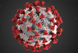
COVID19 (coronavirus disease 2019) is the now well known and infamous infectious
respiratory disease caused by SARS COV 2 virus (Severe Acute Respiratory Syndrome
coronavirus 2). As early as on March 11th,2020 The World Health Organization (WHO) declared COVID 19 a pandemic considering the rapid spread of the disease to multiple countries around the world. To control the spread of COVID 19, heath care authorities in different countries recommended isolation of sick persons, quarantine for those who may been exposed to the virus and social distancing. A distance of at least 6 feet (2 meters) was recommended. Despite these measures rapid spread of the disease and tremendous loss of human lives has occurred worldwide.

Systemic failures led to this enormous and tragic loss of lives. In this presentation, we look at some of these failures and the lessons which can be learnt from them.

When a seizure is not a seizure…let us talk about Pseudoseizures

Nitin K Sethi, MD, MBBS, FAAN

sethinitinmd@hotmail.com

Epilepsy is a common neurological condition in which patients suffer recurrent seizures (also referred to as convulsions).

A generalized convulsion is a rather dramatic event. If it occurs while the patient is standing, frequently the patient gets no warning and falls down striking the ground hard. This is the time injuries occur. As the patient is falling down, he/she is amnestic for the entire episode. Family/bystanders notice that initially the patient stiffens (arms and legs are extended, eyes are rolled up into the head, clenching of the teeth occurs which at times leads to the tongue getting bitten, the patient may at times suffer loss of bladder control). This “tonic” phase is followed by the “clonic” phase during which convulsive movements occur. The seizure stops in a minute or two but the patient remains unresponsive and slowly regains consciousness.

If the above convulsion occurs WHILE the patient is undergoing an EEG test (electroencephalogram), the abnormal brain activity is picked up by the test (see example below) and the diagnosis confirmed.

Based on the misfiring recorded on the EEG, the neurologist can then opine what kind of epilepsy the patient has and where (which part) in the brain the seizures are originating from.

EEG showing abnormal brain waves (spike wave discharges) and beginning of a seizure (IMAGE SOURCE: Wikipedia-the image is used for purely educational purpose)

WHEN A SEIZURE IS NOT A SEIZURE–LET US TALK ABOUT PSEUDOSEIZURES

Pseudoseizure is the term used for events that appear to be epileptic seizures but, in fact, are not. So while the patient may have a dramatic event where in he/she shakes, may roll up the eyes, arch his back, moan, make noises and vocalize, THERE IS NO ABNORMAL EXCESSIVE SYNCHRONOUS CORTICAL ACTIVITY (meaning that on the EEG, the brain waves appear normal without any misfiring).

Pseudoseizures are referred to by various names such as psychogenic non-epileptic events (PNES), non epileptic events (NEE), non epileptic seizures, hysterical seizures.

When pseudoseizures are suspected, a neurologist or epileptologist (epilepsy specialist) shall take a detailed history, may order a MRI scan of the brain and then attempt to capture one of these events on VIDEO-EEG MONITOR (Video-EEG is a special kind of EEG in which simultaneous EEG and video recording of the patient is carried out). The goal is to capture one of the patient’s reported events on video-EEG. If no misfiring of the brain is observed during the event, a diagnosis of pseudoseizures is made.

The non-epileptic (“YOU DO NOT HAVE EPILEPSY” OR “THESE ARE NOT SEIZURES”) nature of the events is then explained to the patient and the family and an attempt is made to try to determine the underlying cause (Why is the patient having these events?). There are many causes of pseudoseizures such as mental stress, sexual or physical abuse, personality disorders, dissociative disorders, affective disorders (mood disorder, anxiety, depression), substance abuse disorder, family conflict, conflict or stress at work, problems in marriage among others.

The treatment is usually a combination of psychotherapy and use of medications (selective serotonin reuptake inhibitors). With support and understanding most patients start to improve and the events either stop completely or become less frequent.

COVID-19 and the Death of a Hospital

Nitin K Sethi, MD, MBBS, FAAN

sethinitinmd@hotmail.com

April 30th, 2010 dawned just like any other April day. As the sun rose along the eastern seaboard, Manhattan came to life with its golden rays. A healthy glow permeated all around. But not all its residents were blessed with this good health. Saint Vincent was sick, terminally sick with multi organ failure. Its corridors once bustling with doctors and nurses in scrubs lay deserted. Its emergency department once full of patients was eerily quiet. Its intensive care units once a melody of ventilator hums and telemetry monitor beeps were quiet as if a great orchestra had played its last composition. Its cafeteria once full of hungry overworked residents had served its last meal. The end had come slowly but now the writing was on the wall. Terminally ill with no hope of a meaningful recovery they said as they went by. The hospital which had once given the gift of life to so many now itself lay on its death bed. Where critically ill patients once received life-saving infusions of antibiotics found itself deserted in its final battle for survival. Saint Vincent found no one to turn to, no one to hold its hand and no one to administer CPR (cardiopulmonary resuscitation). It thought it was too big to fail but no one stepped forward to bail it out. Saint Vincent hospital died on April 30th, 2010.

Image Source: CNN (the picture is purely for information and education purpose)


Saint Vincent’s Hospital or Vinny’s as the residents affectionately referred to it was a large hospital situated in the heart of the West Village. For more than 150 years it served the residents of New York. 1 This is the hospital where the HIV epidemic first exploded in the late 80’s/ early 90s and where the battle against this disease was fought and ultimately won. Vinny was also at the forefront of providing care and comfort when New York City was attacked on September 11th, 2001. If Vinny had not been allowed to die on that fateful day in 2010, it would have certainly opened its wide arms to take care of coronavirus (COVID-19) stricken New Yorkers young and old. Precious lives would have been saved if only Vinny was alive today.

Making sports safer- concussion evaluation, management and return to play decision making for the non-neurologist

Nitin K Sethi, MD, MBBS, FAAN

sethinitinmd@hotmail.com

Background

American football, soccer, rugby, ice hockey, boxing, mixed martial arts, and wrestling are popular contact and combat sports both in the United States and in countries around the world. In these sports head impact exposures resulting in concussions are common. In American football, rugby and soccer, concussive injuries to the brain occur accidently when there is contact of the player’s head with the opposing player(s) head, torso, turf or as occurs in soccer with the ball while heading. In boxing every punch thrown at the head is thrown with the intention of winning by causing a knockout. As a result, concussions, and acute traumatic brain injuries such as subdural and epidural hematoma are common. Subdural hematoma is the most common cause of boxing related mortality. In recent years, the risk of chronic traumatic encephalopathy (CTE), a chronic neurodegenerative disorder associated with contact and combat sports has garnered the attention of physicians, players, and fans.

Concussion definition and evaluation

Concussion is defined as a clinical syndrome characterized by immediate and transient alteration in brain function, including alteration of mental status and level of consciousness, resulting from mechanical force or trauma. It is important to remember that loss of consciousness is not required for a concussion. Concussions are informally and non-scientifically graded into mild, moderate, and severe based on the duration of loss of consciousness and post traumatic amnestic period. Concussive properties of any head impact exposure depend upon the force, velocity, and angle of the hit to the cranium and how the mechanical forces are transferred and absorbed through the intracranial cavity. Both linear and rotational (angular) acceleration forces play a role as does impact deceleration occurring when the athlete falls and strikes the head against the ground.

Image Credit: Patrick J. Lynch, medical illustrator – Modified version of Image: Skull and brain normal human.svg by Patrick J. Lynch, medical illustrator (Source Wikipedia). The image is solely for educational purposes only.

The nature of many contact and combat sports is such that head impact exposures cannot be altogether avoided. Abundant medical literature highlights the long-term health significance of multiple head impact exposures. Minor concussions (sub concussive injuries) contribute to the development of CTE, a neurodegenerative disorder presenting with a constellation of cognitive, mood, and behavioral changes along with motor system dysfunction (Parkinsonism) usually after the athlete has retired. The symptoms of concussion are predominantly subjective such as headache, dizziness, nausea, light sensitivity, sound sensitivity, and cognitive dysfunction. Frequently, when athletes sustain head impact exposures, they themselves are unaware that they are concussed and continue to play. As a result, it is important that when an athlete sustains a head impact exposure, a standardized concussion evaluation be conducted. While this can be conducted on the sidelines, evaluation in the locker room where the athlete is less likely to be distracted is more ideal. Since some athletes exhibit motor signs such as impaired coordination, balance, and stance after a concussion while others exhibit only cognitive dysfunction, it is important that multimodal concussion evaluation be conducted. This includes use of Maddocks questions to gauge awareness, standardized concussion assessment tools such as SCAT5, King Devick test and Balance Error Scoring System (BESS).

Concussions are common in soccer especially during heading the ball.

Management of concussive injury

The athlete’s mentality is to never quit. Hence many athletes will deny symptoms of a concussive injury to the brain. Physicians should be aware of this. An athlete who has suffered a concussion should be pulled out of play and “benched.” This is done for two main reasons. A concussed athlete with impaired attention, concentration, balance, and coordination is more prone to a second head impact exposure. The other reason is that if the concussed athlete continues to play, it is likely that he or she will exhibit more profound and prolonged post-concussion symptoms. The benched athlete is advised a period of cognitive and physical rest. The rationale for this is that a concussed brain is in a state of energy crises and fares better when the cognitive and physical demands on it are less. Cognitive rest entails pulling back from cognitive activities such as team meetings, college work, screen time (amount of time spent using devices with screens such as a smartphone, computer, television, or video game console). Physical rest entails pulling back from normal physical activities such as running and jogging. Here it is important to emphasize that complete cocooning is ill-advised with recent research work showing that it may in fact be detrimental and lead to prolongation of symptoms. The concussed athlete should be evaluated by a physician skilled in concussion management. In most cases neuroimaging with computed tomography or magnetic resonance imaging is not warranted and if conducted comes back negative. There is currently no validated imaging or biofluid (blood or cerebrospinal fluid) biomarkers for concussion. In the absence of biomarkers, the diagnosis of concussion is made clinically based on history of head impact exposure followed by characteristic symptoms (post-concussion symptoms). The concussed athlete is advised to drink adequate water and to avoid alcohol intake and sudden inversions till symptomatic. The role of supplements such as magnesium, vitamin B2 (riboflavin), vitamin B12, fish oil among other remains unclear but these are frequently prescribed. There is no treatment for the head injury itself. Treatments for symptoms of concussion such as headache, dizziness, cognitive impairment, poor sleep, mood, and behavioral disorder involves a multi-disciplinary team consisting of neurologists, neuropsychologists, neurosurgeons, neuroradiologists, neuro-ophthalmologists, physical therapists, occupational therapists, and vestibular therapists with expertise in concussion management.

After a few days of cognitive and physical rest when the acute concussion symptoms have abated, the athlete begins a graded and gradual return to play (RTP) program preferably under the guidance of an athlete trainer with expertise in concussion management. Nowadays most contact and combat sports have sport specific return to play (RTP) protocols. Despite individual variations, the basis tenant remains the same and involves the athlete progressing from light aerobic activity (walking or stationary bike for 10 minutes, no resistance training) to sport-specific activity/drills to non-contact training drills to contact practice and if asymptomatic return to play after a release has been signed by the treating physician.

Conclusion

Concussion is a common head injury in contact and combat sports. Timely identification of the concussed athlete, removal from play and medical management usually results in a good outcome. Closer medical supervision of sports and education of all concerned parties on concussion recognition and management remains the need of the hour.

Facial Nerve Disorders-from Tics to Bells Palsy to Hemifacial Spasm

Nitin K Sethi, MD, MBBS, FAAN

sethinitinmd@hotmail.com

In this blog post, I shall discuss a number of disorders that affect the facial nerve.

The movements of the face are controlled by the facial nerve. This is the nerve which makes us smile, laugh, frown controlling numerous other facial expressions which humans possess. Facial nerve is a cranial nerve (cranial nerves, a set of 12 nerves originate in the brain). Facial nerve is cranial nerve VII (seven). The right facial nerve controls the muscles of facial expression on the right side of the face while the left facial nerve does the same for the left side of the face).

Innervation of the muscles of the face by facial nerve (cranial nerve VII) Henry Vandyke Carter and one more author – Henry Gray (1918) Anatomy of the Human Body. Image source: Facial Nerve Wikipedia. The image is on public domain and is reproduced here for educational purposes only.

There are a number of disorders which can affect the facial nerve. Some of these disorders cause twitching of the face (tics, hemifacial spasm) while others cause weakness/drooping of one side of the face (stroke, Bells palsy).

FACIAL TICS

Facial tics are repeated spasms which involve different muscles of the face (such as rapid eye blinking, nose twitching, head jerking, shoulder shrugs). Tics can be classified into simple motor tics (such as those listed above) and complex motor tics (in a complex motor tic, a series of movements are performed in the same order repeatedly). While tics occur involuntary, most patients are able to stop their tics (for a short period of time) if asked to do so. “Holding the tic in” though causes distress which is relieved by performing the tic. Tics are usually considered to be harmless but they may occur multiple times during the day causing distress and become socially disabling to the patient and those around). As a result, not all tics need to be treated. If the tics are not bothering the patient per se, I usually educate the patient/family on facial tics and what can exacerbate them (stress/anxiety, lack of sleep) and keep the patient under my observation. Many children/ youth may exhibit a transient tic disorder and then grow out of it. Tourette’s Syndrome is a chronic tic disorder characterized by multiple motor and vocal tics. If tics need treatment, behavioral therapy and medications are prescribed.

HEMIFACIAL SPASMS

As the name suggests, in hemifacial spasm the patient exhibits facial muscle contractions (spasms) involving one side of the face. The disorder is characterized as a movement disorder of the facial nerve in which muscles of one side of face twitch involuntary. Hemifacial spasm occurs when something irritates the facial nerve and the cause needs investigation. The diagnosis of HS is made by a neurologist based on observing the facial movements and results of test such as MRI brain (to look and see if anything is irritating the facial nerve/its branches inside the brain).

The treatment of HS depends on the cause. Various treatment options include use of medications (anticonvulsant drugs are sometimes prescribed), botulinum toxin (BOTOX) and surgery.

Treatment of Obstructive Sleep Apnea PART 2

Nitin K Sethi, MD, MBBS, FAAN

You can reach me at: md.nitin@gmail.com

In this blog post, I shall discuss various treatment options for obstructive sleep apnea (OSA). Good news is that nowadays there are a number of very effective treatment options available for OSA. Let us discuss these one by one:

Positional Sleepers: This is the simplest treatment available for snoring and at times OSA. As the name suggests, a positional sleeper helps a person sleep on his/her side as against the back. Since snoring and OSA is frequently worse on the back, sleeping on the side is helpful (the tongue falls on the side and does not obstruct the flow of air). There are many different positional sleepers available on the market. Many of my patients design their own positional sleeper by using pillows or tennis balls sewn into their T shirt/PJs.

COMMERICALLY AVAILABLE POSITIONAL SLEEPER: THIS ONE IS CALLED ZZOMA (Source: sleep education blog) no copyright infringement is intended and the image has been included for purely educational purposes.

Mandibular Advancement Device (MAD): MAD is like a mouth guard. The patient puts the MAD into his/her mouth at night before sleeping. As the name suggests the device “helps to advance the mandible/lower jaw”. The tongue is attached to the lower jaw. So the device by pulling the jaw and tongue forward helps to make more room in the back of the throat for air to flow in and out more easily. There are MADs which are available over the counter. These devices while cheaper as compared to custom made device (made by a dentist) have one problem. They usually are not well fitting. As a result they are not comfortable and are not effective. The best MAD is one which is made by a dentist. The dentist measures the patient’s teeth and makes a device which fits well, is comfortable and effective. Most dentists make MADs so please talk to your dentist about this. There are dentists who are certified by the American Academy of Dental Sleep Medicine. In my opinion these dentists are the best in MADs and I frequently refer my patients to one.

Mandibular Advancement Device (Source: myhealth.alberta.ca) no copyright infringement is intended. Image has been included for purely educational purpose.

Nasal Continuous Positive Airway Pressure (CPAP): Nasal CPAP is the most effective treatment for OSA around the world. CPAP has no side effects-it is a simple machine which helps to “force” the air into the narrowed airway. The patient sleeps with a mask over the nose (nasal mask) or over the nose and mouth (full face mask). The mask is attached to the CPAP machine with the help of a long hose. In the night when the patient is about to sleep, he/she puts the mask on and turns on the machine. The machine sucks in room air, filters it (the machine can be fitted with HEPA filters), humidifies it (the machine has a small humidifier and the patient can change the setting to his/her comfort level) and then blows the air with force in to the patient’s nose. The machine does this throughout the night. In the morning when the patient gets up, the machine is turned off. Indeed a simple and effective solution to OSA. While nasal CPAP has no side-effects, there is one problem. That problem is that majority of patients initially find the CPAP uncomfortable to use. In my experience this is very common. I have patients who come back and tell me they hate the machine, they cannot sleep with the machine and that they will not use it. There are others who come back and say they love the machine and it has changed their lives. So, I tell my patients not to get dejected and upset when initially they are struggling with CPAP use. Finding the most comfortable mask, adjusting the CPAP pressure and humidifier settings wells and most patients over time begin to get used to sleeping with the machine and start to like it. The correct CPAP pressure (the pressure needed to make the OSA go away) is calculated in the Sleep Lab (patient sleeps in the Sleep Lab and while asleep the correct CPAP pressure is determined by the Sleep Medicine technician). There are numerous CPAP machines (different models made by different vendors) and many different types of masks. The trick in my opinion is to find the right mask for the patient and the correct CPAP settings. Once that is done, most patients like CPAP. I usually instruct my patients to try to use the CPAP as much as possible (goal is to get the patient to use the CPAP every night and throughout the night as many patients go to sleep with the CPAP on but then take it off during the night).

Source: CPAP. Wikipedia (no copyright infringement is intended. Image has been included for purely educational purpose).

Pregnancy and epilepsy—when you’re managing both

Nitin K. Sethi, MD; Amy Wasterlain, MD candidate; Cynthia L. Harden, MD

Epilepsy is one of the most common neurological conditions. There is an enormous unmet need when it comes to the care of the epilepsy patient. A few years ago I coauthored an article on the care of pregnant women with epilepsy. I am hopeful that physicians, patients and caregivers shall find the article helpful.

When a patient with epilepsy is pregnant or planning
for pregnancy, you face the challenge of balancing
the benefits and teratogenic risks of her antiseizure
medication. Here’s help.

Here is the link to the full article:

https://www.researchgate.net/publication/49662388_Pregnancy_and_epilepsy-when_you’re_managing_both

Nitin K Sethi, MD, MBBS, FAAN

Epilepsy and Brain Care Center

sethinitinmd@hotmail.com

LET US JOIN HANDS TO DEFEAT EPILEPSY!