


Progressive cerebral atrophy after anoxic encephalopathy following cardiac arrest: a serial MRI study
Prahlad K Sethi and Nitin K Sethi
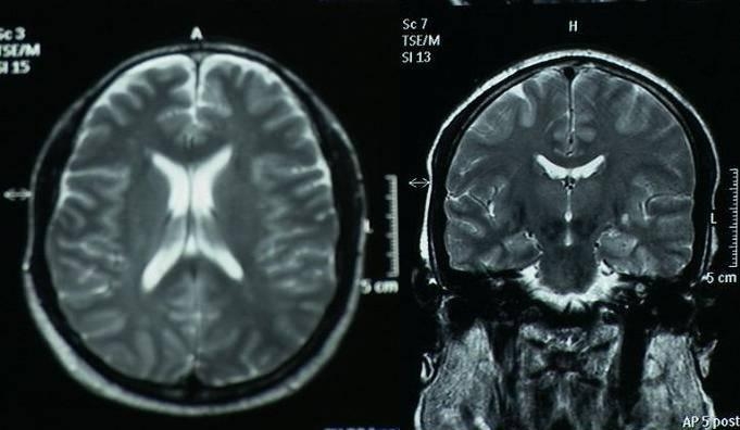
We present serial MRI studies in the case of a 45-year-old man who sustained an in hospital cardiac arrest and was successfully resuscitated after 25 minutes of cardiopulmonary resuscitation. Five years after the cardiac arrest he remains in a persistent vegetative state. Serial MRI studies (Fig 1 immediately and Fig 2 after 3 years and Fig 3 after 5 years of cardiac arrest) indicate ongoing cerebral atrophy and highlight the delayed effects of an initial anoxic injury.
References
1. Cho HJ, Kim HY, So Y. Delayed postanoxic encephalopathy with serial MRI and PET studies. Eur Neurol. 2009; 61(5):315-6.
Legends
Serial MRI axial and coronal sections studies (Fig 1 immediately and Fig 2 after 3 years and Fig 3 after 5 years of cardiac arrest) indicate ongoing cerebral atrophy and highlight the delayed effects of an initial anoxic injury.

I am a 62 year old married white male who had a cardiac arrest 16 years ago. I had the cardiac arrest in a restarant, where I was lucky enough to have three nurses dining as well. According the nurses, they performed CPR for over 10 minutes, that there was at least two or three minutes where there was no pulse detected. After numerous shocks from the paramedics I was brought back to life.
At first, I had severe short term memory problems and a significant hit to my executive functioning skills. Four years later I took a full day of tests for a Neuropsychological Evaluation. My memory was much better, but still had some issues with executive functioning skills. Two years ago I went through another Neuropsychological Evaluation and was told that there continued to be improvement.
Also, about five years ago I had a pet scan (I have now a defibrillatorand cannot have a MRI). One of the doctors later told me that my brain looked similar to an 80 year old and that it was the result of my brain damage at the time of cardia arrest.
This year, I started having mild seizures. EEG test showed that my brain waves behaved very similar to a state of deep sleep. They continue to get worse as time goes by.
My question is one about progressive cerebral atrophy after a cardiac arrest. Is it a resonable assumption that my atrophy continues to worsen and that the seizures are a result of that atrophy?
I am a 29 year old female who in august of 2008 woke up in intensive care. I had suffered an cardiac arrest due to anoxic encephalopathy. I was found not breathing by a friend who then called 911. I was on life support and hospitalized for 60 days and following that I was in rehabilitation for 10 days. When I woke up I had lost all motor skills. I recovered mostly but the doctors have recently told me that I have brain atrophy.